Monday, April 1, 2013
dentalremedies: COSMETIC DENTISTRY { MIDLINE DIASTEMA }
dentalremedies: COSMETIC DENTISTRY { MIDLINE DIASTEMA }: Midline Diastema : The term 'midline diastema' refers to any spacing or gaps existin...
dentalremedies: COSMETIC DENTISTRY { MIDLINE DIASTEMA }
dentalremedies: COSMETIC DENTISTRY { MIDLINE DIASTEMA }: Midline Diastema : The term 'midline diastema' refers to any spacing or gaps existin...
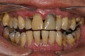
COSMETIC DENTISTRY { MIDLINE DIASTEMA }
Midline Diastema :
The term 'midline diastema' refers to any spacing or gaps existing in the midline of the dental arch. Maxillary midline diastema are one of the most common problems encountered. It has been defined as a space greater than 0.5mm between proximal surfaces of adjacent teeth.

TREATMENT OF MIDLINE DIASTEMA :
1. ORTHODONTIC TREATMENT : Management of Midline Diastema can be done in three phases :-
a) Removal of Cause
b) Active Treatment
c) Retention
a) Removal Of Cause : 1st phase involves the removal of cause. Habbits should be eliminated using Fixed or Removable habbit breaker appliances. In case of imperfect fusion at midline the excision of included interdental tissue between the incisors ( front teeth ) is performed.
b) Active Treatment : This is performed by either by Removable Orthodontic Appliances or Fixed Orthodontic Appliances.

c) Retention : In case of midline diastema long term retention is required. Midline Diastema is considered as easy to treat but difficult to retain.
2. TREATMENT OF MIDLINE DIASTEMA BY COSMETIC RESTORATION :
Esthetic Restorative {filling} Materials i.e. composite resins generally used to close midline diastema specially in adults. It requires a gradual composite build up on the proximal surfaces of Central Incisors and Lateral Incisors in order to achieve a natural shape and size of the teeth.

3. TREATMENT OF MIDLINE DIASTEMA BY PROSTHESIS OR CROWN :
Presence of abnormally shaped teeth require prosthetic rehabilitation. Missing teeth should be replaced with fixed or removable prosthesis.
PORCELAIN LAMINATE VENEERS FOR TREATMENT OF MIDLINE DIASTEMA :
From a cosmetic standpoint, value of the appearance of one's teeth has taken on a greater importance in today's society. Multiple options are available to treat the esthetic problems. Every treatment modality offers some advantages and disadvantages. The use of Porcelain Laminate Veneers to solve the esthetic or functional problems has been shown to be a better management option especially in anterior esthetic zone ( Front Teeth Region ).
.jpg)
.jpg)
The Treatment of Midline Diastema will improve the esthetics of the person. It will help in normal alignment of teeth which will contribute to the oral health but also goes a long way in the overall well being and personality of an individual.
Wednesday, March 6, 2013
dentalremedies: Diabetics and Dental Complications
dentalremedies: Diabetics and Dental Complications: Diabetics increases the risk of severe periodontal disease. Poorly controlled Type 2 Diabetes are more likely to develop pe...
Diabetics and Dental Complications
Diabetics increases the risk of severe periodontal disease. Poorly controlled Type 2 Diabetes are more likely to develop periodontal disease than well controlled diabetics are. Studies conclude that poorly controlled diabetics respond differently to bacterial plaque at the gum line than well controlled diabetics and non-diabetics. Poorly controlled diabetics have more harmful proteins (Cytokines) in their gingival tissue, causing destructive inflammation of the gums. Beneficial proteins (Growth Factors) are reduced interfering with the healing response to infection. Diabetics tend to loose collagen, a protien that supports gums, skin, tendon, cartilage and bone, in their gum tissue thus hastening periodontal destruction. Vascular disorders like reduced circulation in tiny blood vessels in the gums interferes with nutrition and healing in the gum tissues. Young people with Type 1 Diabetes, especially those with poor control are very vulnerable to early-onset periodontal disease as they reach puberty. Diabetics subjects have more deeper pockets between teeth, which indicates moderate to advanced gum disease.
High blood glucose levels help germs to build up on the teeth and gums and make these problems worse leading to loss of teeth. Increased serum Triglyceride levels in uncontrolled diabetics seems to be related to greater attachment loss and probing depths, which are measures of periodontal disease.
The prospects for fighting periodontal disease are excellent as there are many things that diabetic patients can do to stop the process or correct the disease once it starts :
1. Good Blood Glucose Control : The degree of blood glucose control appears to have a direct relationship to the severity of periodontal disease.
2. Oral Hygeine : Dental check up and scaling (if required) at every six months and in case patient has periodontal disease, then check ups should be held every three months.
3. Watch for Warning signs : Bleeding gums while eating , brushing or flossing. Abnormal changes in mouth such as soreness, bright red gums and tenderness. Receeding gums and teeth looking long, chronic bad breath, ill fitting dentures or any other symptoms. White patches on gums indicate fungal infection (Thrush).
4. Avoid smoking, chewing tobacco etc.
5. Keep Cholesterol and Triglyceride level under control.
Thursday, December 23, 2010
dentalremedies: ORAL SUBMUCOUS FIBROSIS
dentalremedies: ORAL SUBMUCOUS FIBROSIS: "Oral submucous fibrosis (or OSF) is a chronic debilitating disease of the oral cavity characterized by inflammation and progressive fibrosis..."
Tuesday, December 21, 2010
ORAL SUBMUCOUS FIBROSIS
Oral submucous fibrosis (or OSF) is a chronic debilitating disease of the oral cavity characterized by inflammation and progressive fibrosis of the submucosal tissues (lamina propria and deeper connective tissues). As the disease progresses, the jaws become rigid to the point that the sufferer is unable to open his mouth.[1][2] The condition is linked to oral cancers and is associated with areca nut chewing, the main component of betel quid. Areca nut or betel quid chewing, a habit similar to tobacco chewing, is practiced predominately in Southeast Asia and India, dating back thousands of years.
SYMPTOMS:
In the initial phase of the disease, the mucosa feels leathery with palpable fibrotic bands. In the advanced stage the oral mucosa loses its resiliency and becomes blanched and stiff. The disease is believed to begin in the posterior part of the oral cavity and gradually spread outward.
Other features of the disease include:
Dried products such as paan masala and gutkha have higher concentrations of areca nut and appear to cause the disease
Biopsy screening is mandatory before treatment. Treatment includes:
The treatment of patients with oral submucous fibrosis depends on the degree of clinical involvement. If the disease is detected at a very early stage, cessation of the habit is sufficient. Most patients with oral submucous fibrosis present with moderate-to-severe disease. Moderate-to-severe oral submucous fibrosis is irreversible. Medical treatment is symptomatic and predominantly aimed at improving mouth movements.Stem cell therapy for oral submucosal fibrosis
Recently scientists have proven that intralesional injection of autologous bone marrow stem cells is a safe and effective treatment modality in oral sub mucosal fibrosis. It has been shown autologous bone marrow stem cell injections induces angiogenesis in the area of lesion which in turn decreases the extent of fibrosis thereby leading to significant increase in mouth opening
SYMPTOMS:
In the initial phase of the disease, the mucosa feels leathery with palpable fibrotic bands. In the advanced stage the oral mucosa loses its resiliency and becomes blanched and stiff. The disease is believed to begin in the posterior part of the oral cavity and gradually spread outward.
Other features of the disease include:
- Pain in the ear or deafness
- Nasal intonation of voice
- Restriction of the movement of the soft palate
- A budlike shrunken uvula
- Thinning and stiffening of the lips
- Pigmentation of the oral mucosa
- Dryness of the mouth and burning sensation
- Decreased mouth opening and tongue protrusion
Dried products such as paan masala and gutkha have higher concentrations of areca nut and appear to cause the disease
- Excessive consumption of red chiles
- Immunological diseases
- Extreme climatic conditions
- Prolonged deficiency to iron and vitamins in the diet
Biopsy screening is mandatory before treatment. Treatment includes:
- Abstention from chewing areca nut (also known as betel nut) and tobacco
- Minimizing consumption of spicy foods, including chiles
- Maintaining proper oral hygiene
- Supplementing the diet with foods rich in vitamins A, B complex, and C and iron
- Employing a dental surgeon to round off sharp teeth and extract third molars
- The prescription of chewable pellets of hydrocortisone (Efcorlin); one pellet to be chewed every three to four hours for three to four weeks
- Forgoing hot fluids like tea, coffee
- Forgoing alcohol
- Submucosal injections of hydrocortisone 100 mg once or twice daily depending upon the severity of the disease for two to three weeks
- Submucosal injections of human chorionic gonadotrophins (Placentrax) 2-3 ml per sitting twice or thrice in a week for three to four weeks
- Surgical treatment is recommended in cases of progressive fibrosis when interincisor distance becomes less than 2 centimetres (0.79 in).(Multiple release incisions deep to mucosa, submucosa and fibrotic tissue and suturing the gap or dehiscence so created by mucosal graft obtained from tongue and Z-plasty. In this procedure multiple deep z-shaped incisions are made into fibrotic tissue and then sutured in a straighter fashion)
- Pentoxifylline (Trental), a methylxanthine derivative that has vasodilating properties and increases mucosal vascularity, is also recommended as an adjunct therapy in the routine management of oral submucous fibrosis.
The treatment of patients with oral submucous fibrosis depends on the degree of clinical involvement. If the disease is detected at a very early stage, cessation of the habit is sufficient. Most patients with oral submucous fibrosis present with moderate-to-severe disease. Moderate-to-severe oral submucous fibrosis is irreversible. Medical treatment is symptomatic and predominantly aimed at improving mouth movements.Stem cell therapy for oral submucosal fibrosis
Recently scientists have proven that intralesional injection of autologous bone marrow stem cells is a safe and effective treatment modality in oral sub mucosal fibrosis. It has been shown autologous bone marrow stem cell injections induces angiogenesis in the area of lesion which in turn decreases the extent of fibrosis thereby leading to significant increase in mouth opening
Subscribe to:
Posts (Atom)